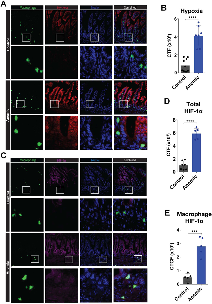
Figure 5. Intestinal macrophages exposed to hypoxia in anemic pups express HIF1α.

(A) Fluorescence microscopy imaging of intestines isolated from control or anemic pups at P8, following administration of Hypoxyprobe (Pimonidazole HCl) 1 hour prior to sacrifice and collection of intestinal tissue followed by staining of frozen sections for macrophages (CD11b) (green), hypoxia (pimonidazole) (red) and nuclei (DAPI) (blue). White boxes indicate enlarged regions directly below each corresponding 20x image. (B) Quantification of hypoxia staining in 5A. (C) Fluorescence microscopy imaging of intestines isolated from control or anemic pups at P8, followed by staining for macrophages (CD11b) (green), HIF1α (magenta) and nuclei (DAPI) (blue). White boxes indicate enlarged regions directly below each corresponding 20x image (D-E) Quantification of total HIF1α staining in 5C (D) and macrophage specific HIF1α staining in 5C (E). CTF = corrected total fluorescence. Data are presented as mean ± SEM, ***P < .001 ****P < .0001.
