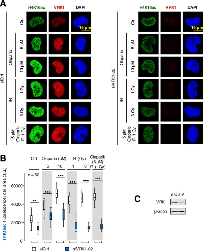
Fig. 2.
Effect of VRK1 depletion on the nuclear fluorescence associated to the acetylation of histone H4 in lysine 16 (H4K16ac ) induced by olaparib, IR or their combination in H1299 (TP53−/−) cells deprived (0.5%) of serum. a left. Effect of si-Control (siCtrl/siC) on H1299 cells treated with different doses of olaparib, IR or their combination on nuclear H4K16 ac fluorescence. a right. Effect of si-VRK1 (siVRK1/siV) on H1299 cells treated with different doses of olaparib, IR or their combination on the acetylation of histone H4 in lysine 16. b. Quantification of the effect of VRK1 depletion on the increase of nuclear H4K16ac fluorescence induced by DNA damage. c. The immunoblot shows the effect of VRK1 depletion on its protein level. siC: siControl, siV: siVRK1–02. ns: not significant, ** p < 0.01, *** p < 0.001. The images show the detail of the subnuclear protein detected. The quantifications were performed using fifty cells from different fields of the experiments (usually between seven and ten were required). The images selected for presentation in the main figure are indicated in Additional file 4: Figure S4. Similar results were obtained in A549 cells (TP53+/+) (Additional file 5: Figure S5) in serum (0.5%) deprived cells

