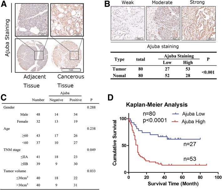
Fig. 1.
High expression of Ajuba predicts poor prognosis in PDAC. a Expression pattern of Ajuba in paired human PDAC specimens (cancerous vs. adjacent tissue) based on intensity and percentage. b Representative images of PDAC tissues with weak/moderate/strong Ajuba expression. Statistics of the IHC staining of 80 cases of the PDAC specimens on TMA chips. Only IHC scores ≥4 point (++) was considered as high. Bar: 20 μm. c High level of Ajuba is positively associated with TNM stage and tumor volumes. d Survival analysis of patients with PDAC by Kaplan–Meier plots and log-rank tests

