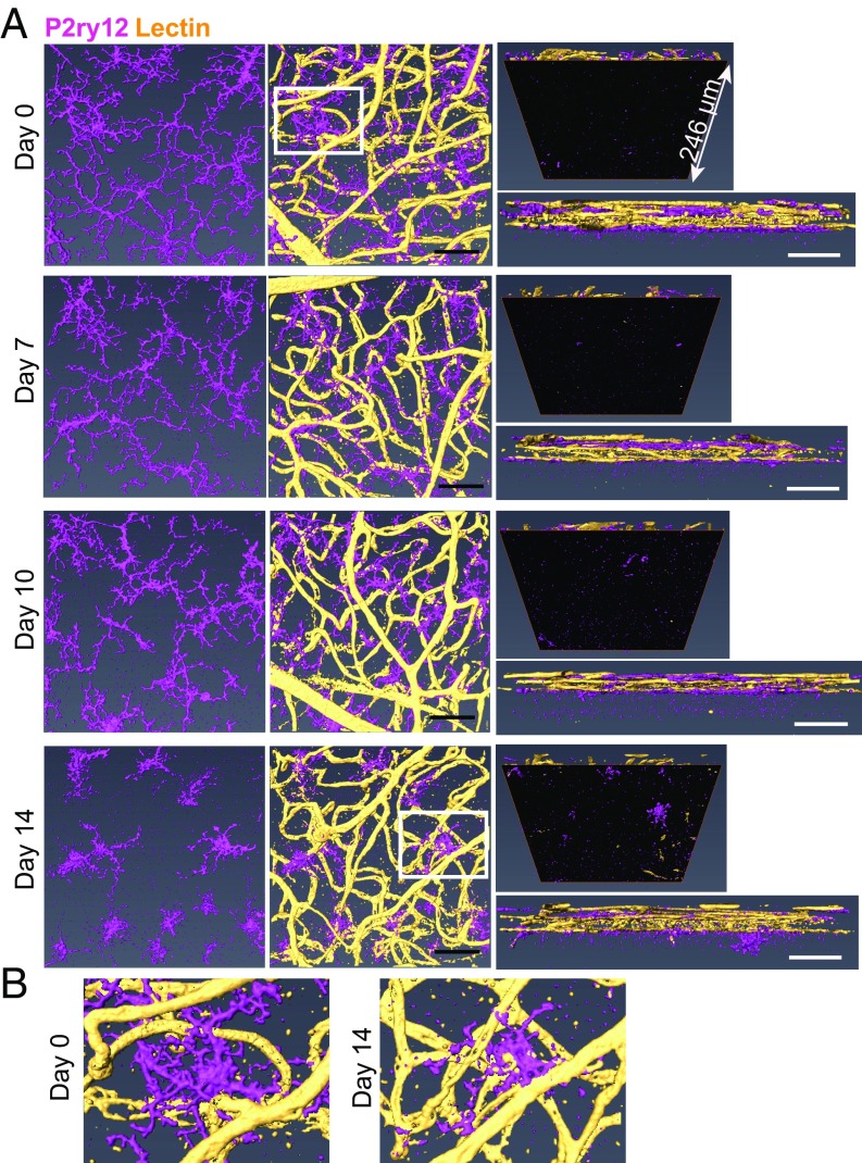
Fig. 6.
Microglia interact with retinal vessels during development of EAU. EAU was induced in C57BL/6 mice, and whole-mount retinas were then stained with anti-P2ry12 antibody (magenta) and lectin (yellow) at 0, 7, 10, and 14 d after EAU induction. Confocal z-stack images of the entire retinal thickness in the midperipheral retina were taken and 3D-reconstructed using Amira software. (A) Representative images from a single scan area at each time point are shown: the top view of microglia (Left), the top view of microglia and lectin (Center), the bottom view (Top Right), and the side view (Bottom Right). The bottom view illustrates changes occurring under the retinal vascular bed (i.e., the outer nuclear layer and the photoreceptors), which are highlighted by masking the upper retina with an inserted black surface. (B) Magnified images of microglia from the areas surrounded by white squares in the images of day 0 and day 14 in A. At least four eyes were examined for each time point. (Scale bars: 50 μm.)

