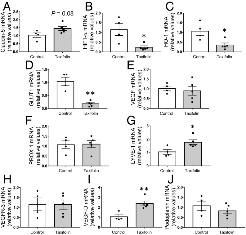
Fig. 2.
Effects of taxifolin on expression levels of blood and lymphatic vasculature-related or hypoxia-responsive genes in the hippocampus of Tg-SwDI mice. Each histogram compares values for the same 14-mo-old Tg-SwDI mice that received either the control diet (n = 4) or taxifolin-containing chow (n = 5) for 13 mo. mRNA levels were analyzed by quantitative RT-PCR and normalized to GAPDH. (A) Expression levels of tight-junction–related cerebrovascular endothelial marker, claudin-5, in the hippocampal tissue. (B–E) mRNA expression levels of hypoxia-responsive genes in the hippocampal tissue: HIF-1α (B), HO-1 (C), GLUT1 (D), and VEGF (E). (F–J) Gene mRNA expression levels of markers for lymphatic endothelial cells in the hippocampal tissue: PROX-1 (F), LYVE-1 (G), VEGFR-3 (H), VEGF-D (I), and podoplanin (J). Data are expressed as mean ± SEM (control, n = 4; taxifolin, n = 5). P values were determined by Student’s t test. *P < 0.05; **P < 0.01.

