Fig. 2.
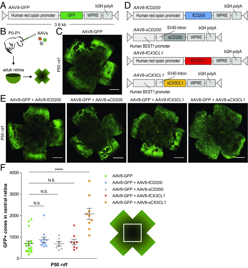
Effect of CD200 and CX3CL1 overexpression on cone survival. (A and B) Schematics of AAV8-GFP vector and delivery. (C) Flat-mounted P50 rd1 retina infected at P0–P1 with AAV8-GFP. (Scale bar: 1 mm.) (D) Schematics of CD200 and CX3CL1 AAV vectors. (E) Flat-mounted P50 rd1 retinas infected at P0–P1 with the indicated AAVs. (Scale bars: 1 mm.) (F) Quantification of cone survival in the central retina of P50 rd1 retinas infected with the indicated AAVs. Data are shown as mean ± SEM (n = 7–18 animals per condition). ****P < 0.0001 by two-tailed Student’s t test with Bonferroni correction. N.S., not significant.

