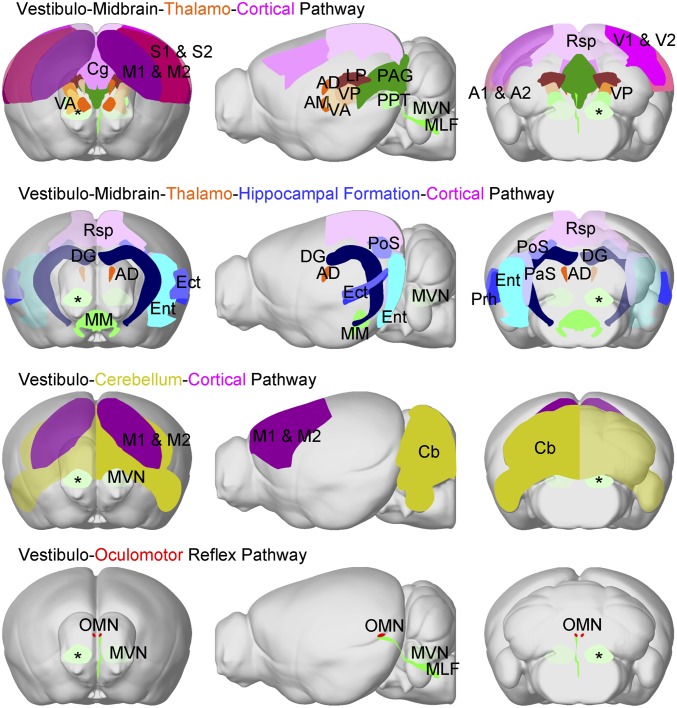
Fig. 5.
Categorization of distinct brain-wide central vestibular pathways activated by optogenetic stimulation of MVN excitatory neurons. We categorized four distinct pathways based on the activated regions detected by BOLD fMRI in Fig. 2. Note that the color used to define each region is scaled to the mean activation strength as quantified by the averaged area under the BOLD signal profile in Fig. 2C. The vestibulo-midbrain-thalamo-cortical pathway and ipsilateral VA and VP showed stronger activations than their contralateral counterparts. This indicates that monosynaptic projections from the site of stimulation in the ipsilateral MVN (*) directly drove activation in the motor and somatosensory thalamus. In the vestibulo-midbrain-thalamo-hippocampal formation cortical and vestibulo-midbrain-hippocampal formation pathways, the majority of hippocampal formation regions (PaS, PoS, Ect, Ent, and DG), a cortical region (Rsp), and a thalamic region (AD) displayed stronger contralateral activations. These regions participate in processing head direction signals (3, 5). The vestibulo-cerebellum-cortical pathway displayed stronger activations at the contralateral Cb and M1 & M2, indicating key interactions underlying vestibulo-motor functions. In the vestibulo-oculomotor reflex pathway, both activated OMNs did not show hemispheric differences despite only receiving direct projections contralaterally from the MVN. AM, anteromedial thalamus.

