Fig. 2.
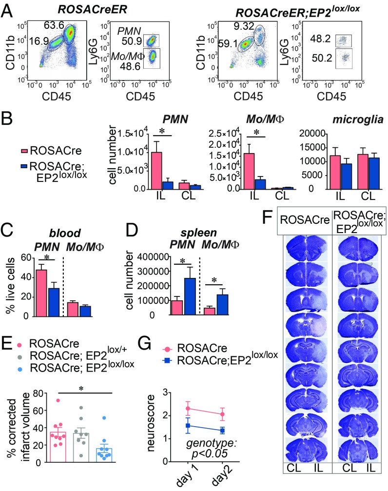
Inducible global deletion of the EP2 receptor reduces myeloid cell infiltration after MCAo and prevents cerebral injury. RosaCreER and RosaCreER;EP2lox/lox 3–4 mo C57B6/J male mice underwent MCAo followed by 48 h RP. Data are presented as mean ± SEM. (A) Representative plots of CD11b+CD45hiLy6Ghi PMN and CD11b+CD45hiLy6Glo Mo/MΦ from RosaCreER and RosaCreER;EP2lox/lox ischemic hemispheres 2 d after MCAo. (B) Cell numbers in IL and CL hemispheres for PMN, Mo/MΦ subsets, and microglia (n = 5–11 per group; two-way ANOVA; for PMN, effect of the genotype, P = 0.026, effect of the hemisphere, P = 0.02; for Mo/MΦ, effect of the genotype P = 0.017, effect of the hemisphere, P = 0.002; Tukey post hoc, *P < 0.05). (C) At 48 h, numbers of PMN and Mo/MΦ in the spleen (n = 5–9 per group; two-tailed Student’s t test, *P < 0.05). (D) At 48 h, percentage of live cells representing PMN and Mo/MΦ in blood 48 h after MCAo (n = 7–10 per group; two-tailed Student’s t test, *P < 0.05). (E) Neurological scores (n = 7 to 8 per group; repeated measure two-way ANOVA, effect of the genotype P = 0.0492; effect of time P < 0.0001). (F) Representative series of brain sections stained with Cresyl violet (CV) from RosaCreER and RosaCreER;EP2lox/lox mice 48 h after MCAo. Areas lacking CV staining were quantified for infarct volume. (G) Quantification of the percentage of corrected hemispheric infarct volume at 48 h after MCAo (n = 8 to 9 per group; one-way ANOVA P = 0.0328; Tukey’s post hoc *P < 0.05).

