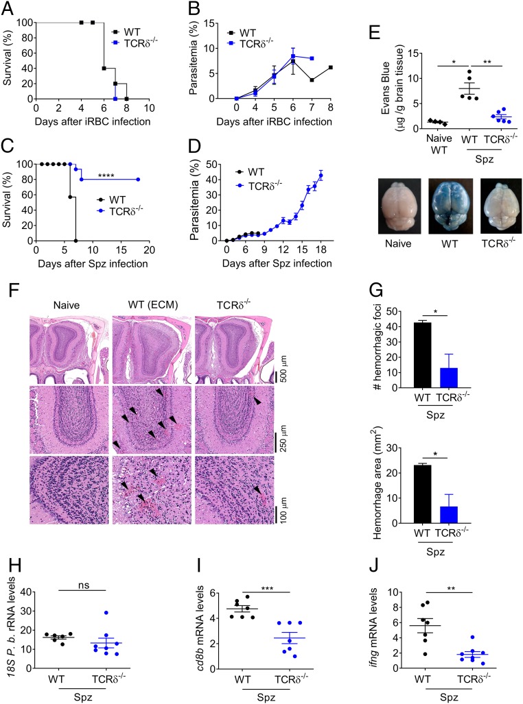
Fig. 1.
γδ-T cells are essential for liver stage-dependent ECM pathogenesis. (A and C) Survival and (B and D) parasitemia curves for WT and TCRδ−/− mice. WT (n = 5–14) and TCRδ−/− (n = 5–15) mice were infected with 1 × 10⁶ P. berghei ANKA GFP iRBCs by i.p. injection (A and B); or 2 × 10⁴ P. berghei ANKA GFP Spz by i.v. injection (C and D). (C) Data combined from three independent experiments [****P < 0.0001 by Log-rank (Mantel–Cox) test]. (E) Assessment of BBB disruption by EB staining of noninfected (naïve, n = 5) versus Spz-infected WT (n = 5) or TCRδ−/− (n = 6) mice. (F) Representative microphotographs of coronal sections of mouse brain at the level of the olfactory bulb by H&E. Original magnification, 2.5×, 10×, and 20×. (Scale bars, 100–500 μm.) Images are representative of three mice. (G) Number and area of hemorrhage foci (n = 3) in the olfactory bulb (approximately at bregma 3.92). (H) Relative expression levels of parasite 18S P.b. rRNA, (I) cd8b, and (J) ifng in brain tissue of WT and TCRδ−/− (n = 6–8) Spz-infected mice (arbitrary units normalized to the housekeeping gene hprt). Data are represented as mean ± SEM. Each symbol in E and H–J represents an individual mouse. *P < 0.05; **P < 0.01; ***P < 0.001; ****P < 0.0001; ns, not significant; by Mann–Whitney U test.

