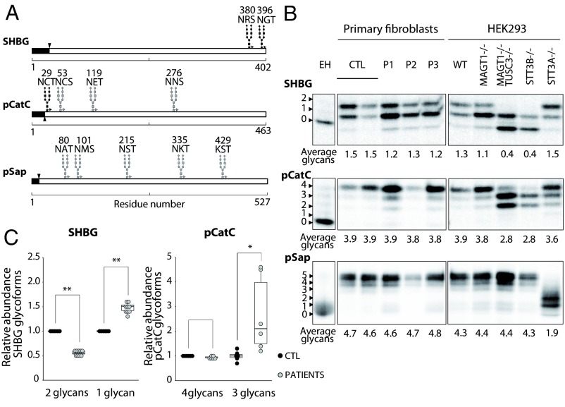
Fig. 3.
STT3B-dependent glycosylation is affected in patients’ fibroblasts. (A) Diagrams showing the glycosylation sites of SHBG, pCatC, and pSap. Black glycan structures indicate an STT3B-dependent site. Signal sequences are depicted in black. (B) HEK293 cells and fibroblasts were transfected with SHBG, pCatC, or pSap, followed by pulse-chase labeling. Quantified values are shown below gel lanes and represent the average number of glycans for the respective reporter (n = 3). (C) Quantification of the different glycoforms of SHBG and pCatC in fibroblasts, normalized to the averaged control samples. EH indicates endoglycosidase H treatment. *P < 0.5; **P < 0.005.

