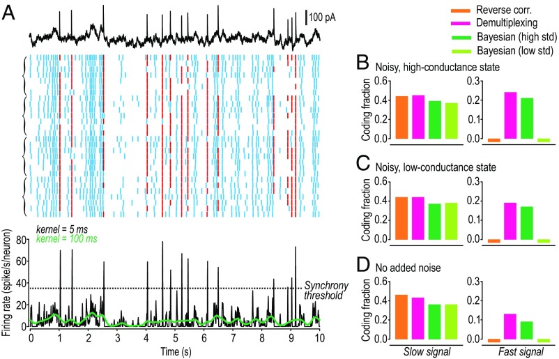
Fig. 4.
Pyramidal neurons can multiplex in vitro. (A) Sample rasters from short length of 100-s-long responses. All neurons received the same mixed signal but different conductance noise on each trial. Four neurons were tested with 7 trials each; brackets on left group responses by neuron. Black FRH was thresholded to identify synchronous (red) or asynchronous (blue) spikes. (B) Decoding of the mixed signal from ensemble response (28 trials) illustrated in A, based on neurons tested in the noisy, high-conductance state. Same analysis conducted on neurons tested in a noisy, low-conductance state (31 trials from six neurons) (C) or in neurons without any added noise (29 trials from five neurons) (D). For all different conditions, the four decoding strategies yielded a pattern of CF values very similar to that seen in simulations (Fig. 3D).

