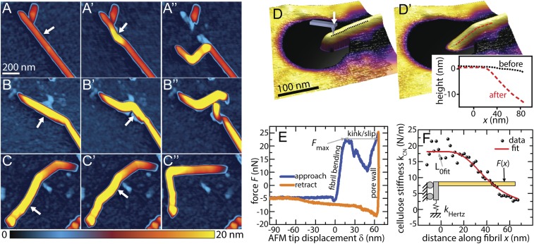
Fig. 1.
Probing mechanical deformation at the nanoscale reveals reversible and irreversible nature of cellulose kinks. (A–D′) AFM images of cellulose nanofibrils before and after a series of lithographic lateral manipulations with the AFM tip demonstrate three typical responses. The arrow in each image indicates the location and direction of applied force. (A–A′′) Multiple kinks were induced along the length of the fibril. (B–B′′) Complete breakage of the fibril was achieved by applying manipulation to the vicinity of an existing kink defect, while the fibril was strongly adhered to the substrate. (C–C′′) A previously kinked fibril was straightened, indicating that the kinks observed in C and C′ still maintain some degree of molecular connectivity. (D) A cellulose nanofibril suspended over a 200-nm pore in track-etched polycarbonate (TEPC). (D′) Kink defect formed in the same nanofibril shown in D after AFM indentation at 48-nN applied load. (Inset) Comparison of the topographic line profile of the nanofibril before and after indentation, clearly revealing the presence of a mechanically induced kink at the supporting pore wall. (E) Approach and retract indentation curves for the particular indentation step that induced kinking in the nanofibril. After reaching the maximum force Fmax in the approach curve the cellulose forms a kink, allowing the probe tip to slip into the pore wall. (F) AFM indentation and bending measurements along the length of a nanofibril were fit to a beam model to calculate deformation stress.

