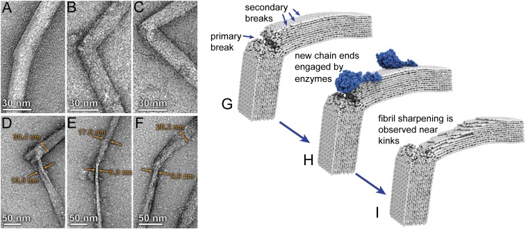
Fig. 3.
Imaging of cellulose nanofibrils before and after enzymatic hydrolysis shows that Cel7A preferentially initiates hydrolysis at the location of kink defects: (A–C) TEM micrographs showing examples of kink defects in isolated Cladophora cellulose nanofibrils. (D–F) After incubation with purified Cel7A, the nanofibrils exhibited localized narrowing in close proximity to the kink defect. Measurements of fibril width indicate that narrowing is more severe on one side of the defect. (G–I) Schematic depiction of the proposed mechanism for localized hydrolysis at kink defects by the processive cellobiohydrolase Cel7A, wherein reducing ends formed by bond breakages at the kink location (G) are engaged by the enzyme (H). Unidirectional hydrolysis results in formation of additional reducing ends on the same side of the kink defect, which are subsequently engaged by additional enzymes. This process results in preferential narrowing of the fibril on one side of the defect as enzymatic digestion progresses (I).

