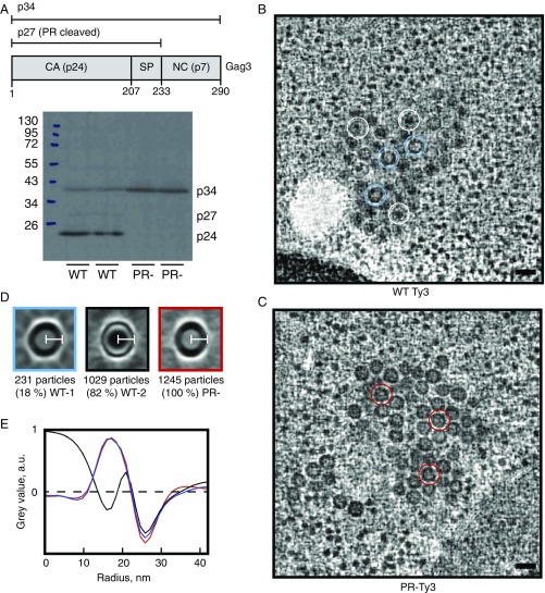
Fig. 1.
WT and PR- Ty3 particle morphology. (A) Schematic Ty3 Gag3 polyprotein showing regions corresponding to p34 (aa 1–290), p27 (aa 1–233), and CA p24 (aa 1–207). Western blot analysis of yeast cells expressing WT or PR- Ty3. Note that Gag3 (p34) and its PR cleavage products (p27, p24) are known to migrate anomalously (41). (B) Slices through representative tomographic reconstructions of resin-embedded yeast cells containing WT Ty3 particles. Representative WT type 1 particles (thick-ring morphology) are marked with blue circles, and representative WT type 2 particles (thin ring morphology) are marked with white circles. (Scale bar, 50 nm.) (C) Slices through representative tomographic reconstructions of plastic-embedded yeast cells containing PR- Ty3 particles. Representative PR- particles are marked with red circles. Particles are homogeneous, and all have a thick-ring morphology. (Scale bar, 50 nm.) (D) Central slices through the particle averages for WT-1, WT-2, and PR- Ty3 populations. (White scale bars, 21 nm.) (E) Radial profiles through the particle averages. The WT-1 and PR- particles with immature-like morphology have the same radius and radial profile, while WT-2 particles with mature-like morphology have the same radius but a different radial profile.

