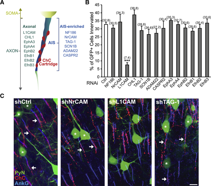
Figure 2. RNAi Screen of PyN-expressed Cell Surface Molecules Identifies L1CAM as a Regulator of Neocortical ChC/PyN AIS Innervation.
(A) Schematic depicting the subcellular localization (i.e. axonal or AIS-enriched) of screened cell surface molecules in neocortical PyNs.
(B) In vivo RNAi screen of PyN-expressed axonal and AIS-enriched cell adhesion molecules and select members of the Eph and ephrin family of receptors/ligands present in the neocortex. Quantification of the percentage of GFP+ PyNs innervated by single RFP+ ChCs at P28 in LII of somatosensory cortex from Nkx2.1-CreER;Ai9 mice electroporated at E15.5 with plasmids expressing EGFP and shRNAs targeting indicated cell surface molecules. Innervation percentages are indicated for each condition. 6–16 ChCs and 12–99 GFP+ PyNs per ChC from 3 animals were analyzed for each condition; one-way ANOVA, post hoc Tukey-Kramer test.
(C) Representative images of PyNs innervated by ChC cartridges in LII of somatosensory cortex from Nkx2.1-CreER;Ai9 mice electroporated at E15.5 with plasmids expressing EGFP and shCtrl, shNrCAM, shLICAM, or shTAG-1 and sacrificed at P28. Scale bar, 10 μm.
***p < 0.001. Data are mean ± SEM. See also Fig. S1 and Table S1.

