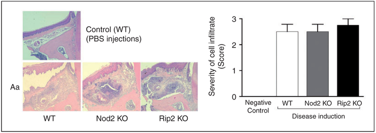
Figure 2.
Inflammatory infiltrate associated with experimental periodontitis in NOD2-KO and RIP2-KO mice. Representative images of 5 μm hematoxylin and eosin-stained sections from upper first molars (frontal or buccal–palatal plane) subjected to Aa injections, according to the genotype of the animal. A representative image of a control mouse (WT, PBS-injected) is shown for comparison purposes (no difference was noted in comparison with PBS-injected tissues of NOD2-KO and RIP2-KO mice; data not shown). The graph presents the analysis of cellular infiltrate using the severity score. At least four semi-serial sections obtained from samples from at least three different animals were analyzed for each group and these representative images were obtained at 100× magnification.

