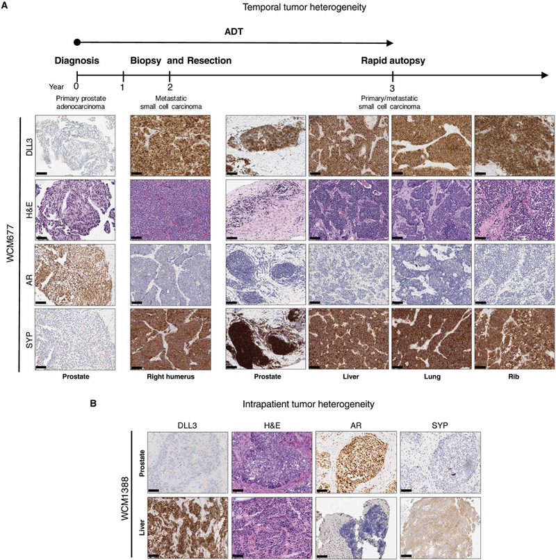
Fig. 2. Temporal and intrapatient tumor heterogeneity in two patients.

(A) Representative H&E histology images with DLL3, AR, and SYP IHC of a patient with prostate cancer, followed from initial diagnosis of prostate cancer (year 0) to autopsy (year 3). Prostate (year 0), right humerus (year 2), and autopsy (year 3). Scale bars, 100 μm. Samples were obtained from the prostate, liver, lung, and rib. Androgen deprivation therapy (ADT) treatment is indicated by the arrow. (B) Representative H&E and representative DLL3, AR, and SYP IHC images of prostate and concurrent liver biopsies derived from the same patient at the same time point. Scale bars, 100 μm.
