
Fig. 5. Clinical response in a patient enrolled on a phase 1 SC16LD6.5 basket trial.
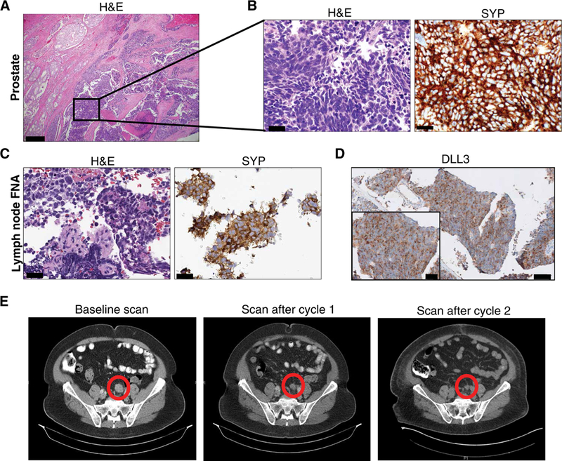
(A) Representative H&E image of radical prostatectomy specimen that shows a PCA (left) with an adjacent component of small cell neuroendocrine carcinoma (inset). Scale bar, 200 μm. (B) H&E and strong diffuse synaptophysin (SYP) IHC of the small cell carcinoma component. Scale bars, 50 μm. (C) Lymph node biopsy performed 2 years after prostatectomy. Representative images of H&E and SYP IHC are shown. Scale bars, 50 μm. Fine-needle aspiration, FNA. (D) Representative images of DLL3 IHC are shown (scale bar, 200 μm; inset scale bar, 50 μm). (E) CT scans before therapy and after cycles 1 and 2 of SC16LD6.5 (nodal metastasis highlighted by a circle).