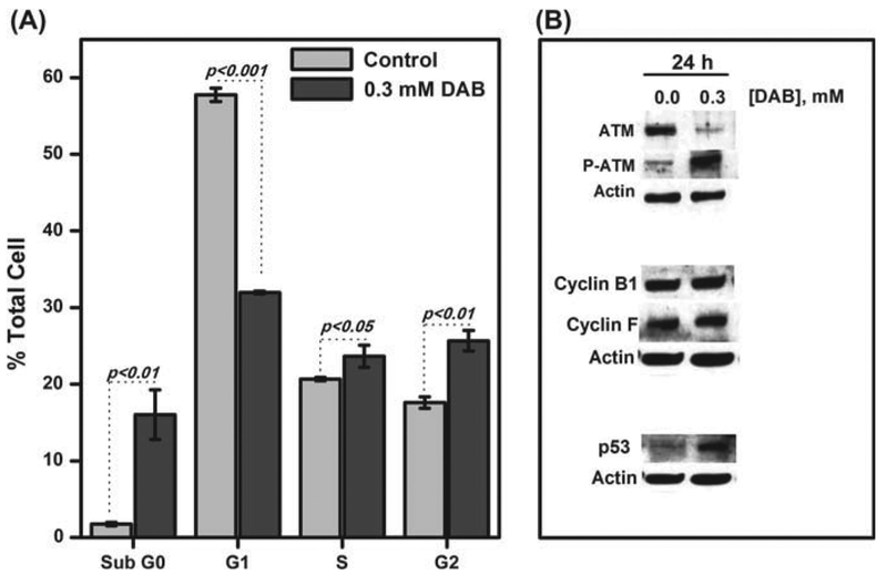
Figure 8. Effect of DAB treatment on cell cycle and related proteins.
RKO cells are treated with 0.30 mM DAB for 24 h in OptiMEM. After time-lapse incubation, the cell suspensions (2 × 10⁶ cells/ml) are fixed in cold 70% ethanol overnight at 4°C, washed with PBS and stained with 50 μ g/mL iodide propidium solution in DPBS containing 50 μg/mL of RNase A for 30 min. The samples are analyzed by a flow cytometer (A). Protein samples are submitted to Western blotting analysis in which specific primary antibodies are used (B).

