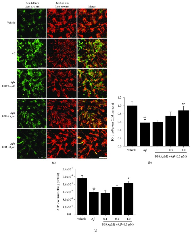
Figure 2.
Berberine ameliorates Aβ1-42-induced changes in mitochondrial membrane potential and the decline of ATP levels in primary cultured hippocampal neurons. (a) Representative images of JC-1 staining of hippocampal neurons treated with vehicle, oligomeric Aβ1-42 (0.5 μM), or oligomeric Aβ1-42+berberine (0.1, 0.3, or 1 μM). JC-1 aggregates (red) indicate healthy mitochondria, while green fluorescence indicates cytosolic JC-1 monomers. Scale bar = 50 μm. (b) The ratio of red to green fluorescence in A was quantified to measure changes in the mitochondrial membrane potential. (c) ATP levels treated with vehicle, oligomeric Aβ1-42 (0.5 μM), or oligomeric Aβ1-42+berberine (0.1, 0.3, or 1 μM). Values are expressed as the mean ± standard error of the mean of 4 independent experiments. ∗∗p < 0.01 vs. the vehicle-treated group, #p < 0.05, ## p < 0.01 vs. the Aβ1-42-treated group. Aβ: cells were treated with Aβ1-42 at 0.5 μM.

