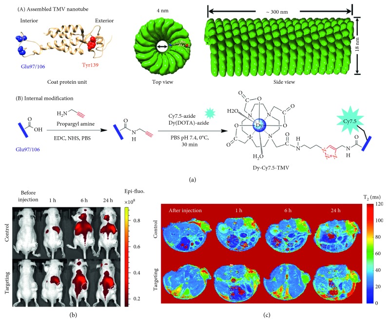
Figure 5.
(a) Structure of the tobacco mosaic virus (TMV) nanoparticle's coat protein with surface-exposed residues highlighted as internal glutamic acid (blue) and external tyrosine (red) and the structure of the assembled capsid/strategy for internal modification. (b) Near-infrared fluorescence (NIRF) imaging of subcutaneous PC-3 (α2β1) prostate tumours in athymic nude mice (n = 3) before and 1, 6, and 24 h after the intravenous injection of Dy-Cy7.5-TMV-mPEG (control group) or Dy-Cy7.5-TMV-DGEA (targeting group). (c) In vivo T2-mapping MRI of subcutaneous PC-3 (α2β1) prostate tumours in athymic nude mice (n = 3) obtained before and 1, 6, and 24 h after the intravenous injection of Dy-Cy7.5-TMV-mPEG (control group) and Dy-Cy7.5-TMV-DGEA (targeting group) (reprinted (adapted) with permission from [62]).

