Figure 2.
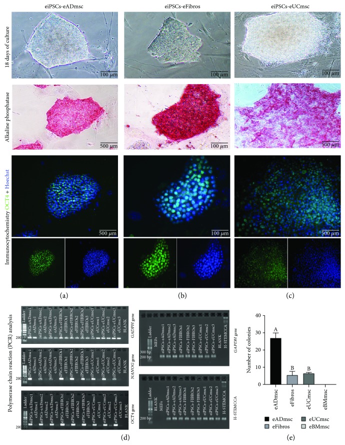
Equine iPSCs on day 18 after transduction from (a) adipose tissue mesenchymal cells, (b) fibroblasts, and (c) umbilical cord tissue mesenchymal cells, 200x. Alkaline phosphatase-positive equine iPSC colonies were induced from each cell type: (a) adipose tissue mesenchymal cells, 100x; (b) fibroblasts, 200x; (c) umbilical cord tissue mesenchymal cells, 100x. In addition, images present the immunocytochemistry expression of merged OCT4, OCT4/FITC, and Hoechst staining: (a) adipose tissue mesenchymal cells, 100x; (b) fibroblasts, 200x; (c) umbilical cord tissue mesenchymal cells, 100x. (d) Transcript expression of GAPDH, NANOG, and OCT4 in equine adipose tissue mesenchymal cells, umbilical cord tissue mesenchymal cells, and fibroblasts before and after pluripotency induction. NANOG and OCT4 expression levels are enhanced after cell reprogramming. Also in (d), confirmation of GAPDH and STEMCCA expression in equine iPSCs by conventional PCR. (e) Graph showing the total production of eiPSC colonies using hOSKM. No colonies were formed from the bone marrow mesenchymal cells. Different letters indicate significantly different results (P < 0.05).

