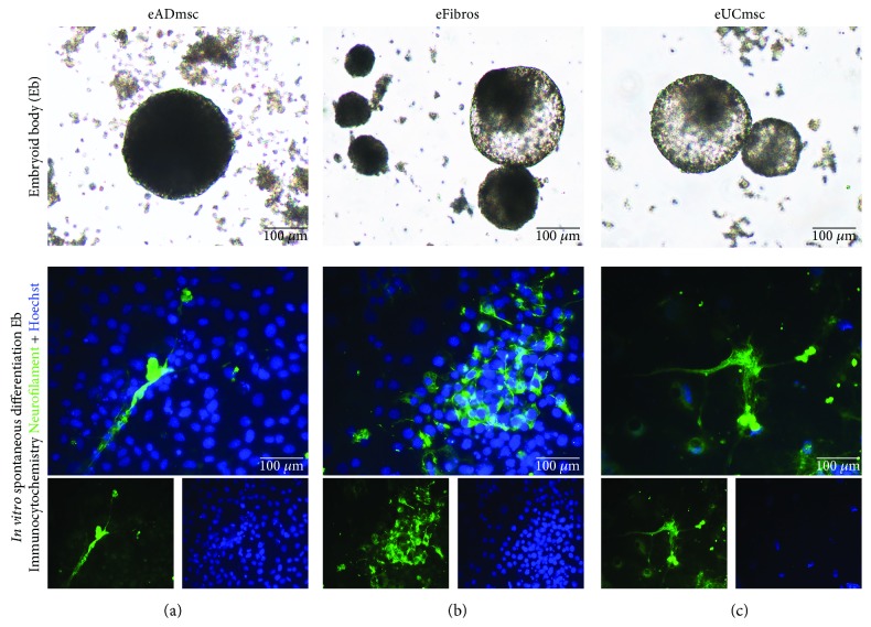
Figure 3.
Four-day-old EBs produced from equine iPSCs derived from (a) adipose tissue mesenchymal cells, (b) fibroblasts, and (c) umbilical cord tissue mesenchymal cells. After the spontaneous differentiation of embryoid bodies into multiple lineages, the cells presented a more elongated morphology and were immunocytochemically positive for neurofilament. Merged images of neurofilament/FITC and Hoechst staining: (a) eiPSCs-eADmsc-derived cells, (b) eiPSCs-eFibros-derived cells, and (c) eiPSCs-UCmsc-derived cells, 200x.

