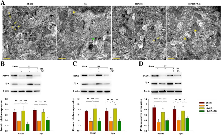
Fig. 5.
Effects of HS on structural remodeling of synapses following HI injury. a Representative electron micrographs at 28 days following HI indicating: * - presynaptic vesicles, Yellow arrow - postsynaptic partners, Green arrowhead - widened synaptic clefts, and Yellow arrowhead - destroyed synapses. Scale bar = 2 μm. b-d Levels of PSD95 and Syn within the ipsilateral cortex as examined at 3, 14 or 28 days post-HI with use of Western blot. Bar graphs show quantifications of protein levels at 3, 14 or 28 days. N = 4/group. Values represent the mean ± SD, * p < 0.05, **p < 0.01, ***p < 0.001 according to ANOVA

