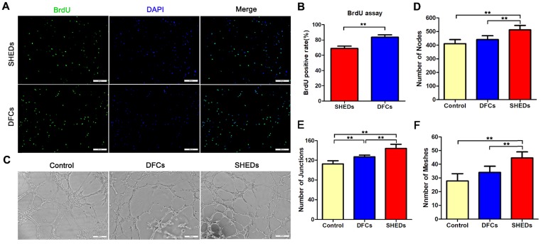
Fig 3.
BrdU incorporation and tubule formation assays using DFCs and SHEDs. Immunofluorescence staining after an incubation with BrdU for 48 h (A). More DFCs displayed BrdU-positive staining, suggesting a higher proliferation rate than SHEDs (B). In vitro tube formation by HUVECs in Matrigel induced by conditioned media from DFCs and SHEDs (C). Typical tube-like structures in different groups are shown. The numbers of nodes (D), junctions (E) and meshes (F) per field of view in the three groups were analyzed. Scale bars =200 μm, * p<0.05 and ** p<0.01.

