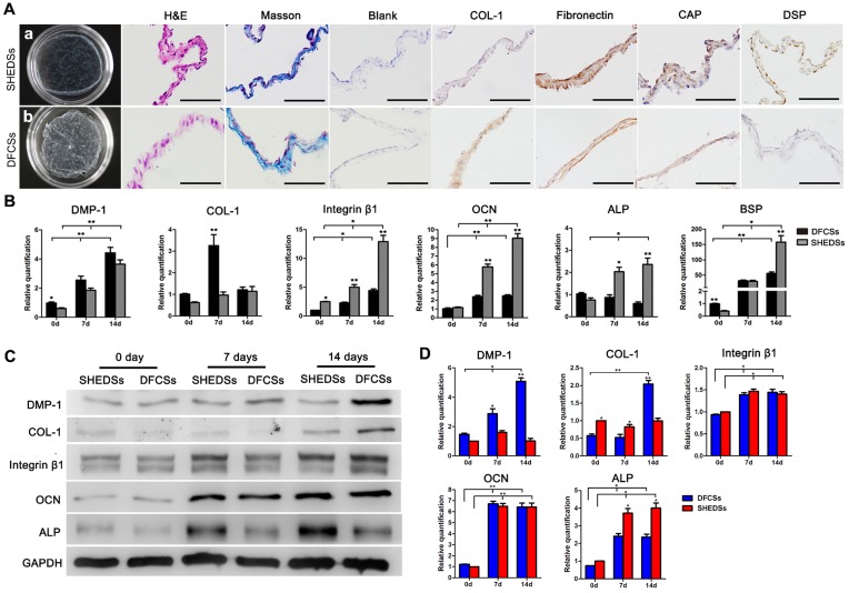
Fig 6.
Cell sheet characteristics and in vitro differentiation of both cell types induced by ECM. Macroscopic view of the cell sheets after culture for 15 days in vitro (Aa and b). HE staining showed that both sheets consisted of 2-3 layers of cells and Masson's trichrome staining indicated that the membranes were rich in collagen fibers (A). Immunocytochemical staining revealed the expression of the ECM proteins COL-1 and fibronectin and odontogenic proteins CAP and DSP in both DFCSs and SHEDSs (A). The expression of odontogenic genes in DFCSs and SHEDSs after ascorbic acid induction for 0, 7 and 14 days was detected using real-time PCR, and relative quantification (RQ) values were calculated (B). Western blotting was also utilized to detect the levels of related proteins, with GAPDH serving as internal control (C) and the relative levels were quantified (D). After forming the cell sheets, DFCSs expressed the DMP-1 and COL-1 proteins at higher levels, while SHEDSs expressed ALP at higher levels. The levels of the OCN and Integrin β1 proteins were not statistically significantly different between the two groups from days 7 to 14. Scale bars =200 μm, * p<0.05 and ** p<0.01.

