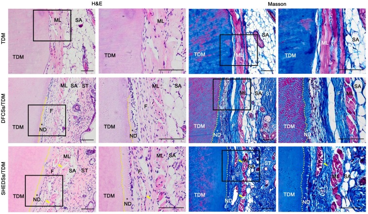
Fig 7.
Odontogenic differentiation of DFCSs and SHEDSs combined with human TDM in nude mice. The composites were transplanted into subcutaneous sites and allowed to grow for 8 weeks. No new tissue was observed in the control group, and the TDM was directly covered by the subcutaneous muscle layer and its fascia. However, newly formed predentin and periodontal ligament-like fibers between the TDM surface and mouse subcutaneous muscle layer were successfully generated in both the DFCSs/TDM and SHEDSs/TDM groups. Masson staining also further confirmed the same results. Scale bars =100 μm. (F: periodontal ligament-like fibers; ML: muscle layer; ND: new dentin; SA: subcutaneous adipose tissue; ST: skin tissue; TDM: treated dentin matrix; yellow arrow: blood vessel).

