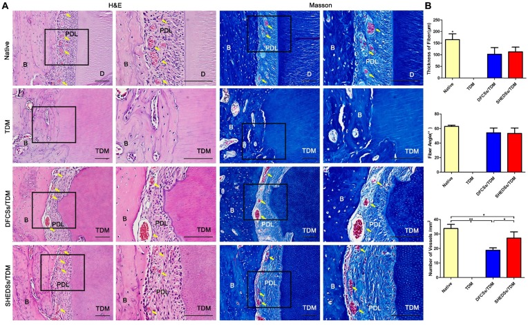
Fig 8.
Orthotopic implantation of bio-root composites in the jaw bones of Sprague-Dawley rats for 8 weeks. H&E staining verified that both the SHEDSs/TDM and DFCSs/TDM groups possessed a clearance between the TDM and jaw bone, which exhibited similar characteristics to native tooth root, including dense collagen fibers, fibroblasts, and blood vessels (yellow arrow) contributing to the formation of periodontal ligament tissues (A). However, in the TDM alone group, neither clearance nor periodontal ligament-like structures were observed between the TDM surface and jaw bone. Masson's trichrome staining also further confirmed the same results. The sizes of regenerated periodontal tissues in vivo were measured (B), including the fiber thickness, the fiber angle and the number of vessels in four groups. Scale bars =100 μm. (B: jaw bone; D: dentin; PDL: periodontal ligament tissue; TDM: treated dentin matrix).

