Figure 6.
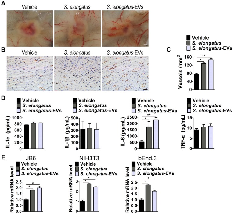
S. elongatus-EVs enhance angiogenesis in the wound sites and IL-6 expression in vivo and in vitro. (A) Gross view of wounds treated with vehicle (PBS), S. elongatus and S. elongatus-EVs at day 12 post-wounding from the undersurface. Scale bar: 2 mm. (B-C) Representative images (B) and quantification (C) of CD31-stained blood vessels in the wound sites. Scale bar: 50 μm. n = 3 per group. (D) ELISA of the concentrations of IL-1α, IL-1β, IL-6 and TNF-α in skin tissue homogenate from mice treated with vehicle, S. elongatus and S. elongatus-EVs at day 7 post-wounding. n = 3 per group. (E) qRT-PCR analysis of the expression levels of IL-6 in mouse epidermal JB6 cells, NIH3T3 fibroblasts and bEnd.3 endothelial cells receiving different treatments for 24 h. n = 3 per group. Data are plotted as mean ± SD. *P < 0.05, **P < 0.01, ***P < 0.001.

