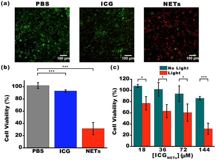
Figure 4.
Photodestruction of SKBR3 cancer cells in vitro. (a) Fluorescent images of SKBR3 cancer cells incubated with PBS (negative control), 176 μM free ICG (positive control), and NETs ([ICGNETs] ≈ 144 μM) for 3 h at 37°C and followed by 808 nm laser irradiation at I0 = 680 mW/cm2 for 15 min. Live cells were identified by the Calcein AM stain and falsely colored in green. Dead cells in response to laser irradiation were identified using the EthD-1 stain and falsely colored in red. (b) Percentage viability of SKBR3 cells as a function of incubation agent. (c) Percentage viability of SKBR3 cells as a function of [ICGNETs]. In panels (b) and (c), statistically significant differences are indicated as *p < 0.05 and ***p < 0.001 (n = 3 samples for each treatment).

