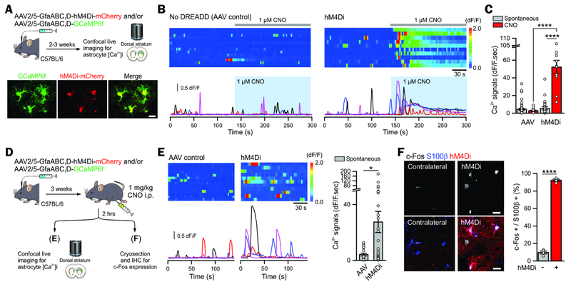
Figure 2: Astrocyte-specific Gi pathway activation by a Gi-DREADD hM4Di.
(A) Cartoon illustrating AAVs used for expressing GCaMP6f with and without mCherry-fused hM4Di in astrocytes in the dorsal striatum. The lower images show GCaMP6f and hM4Di-mCherry expressing astrocytes in striatal slices were colocalised (Chai et al., 2017).
(B-C) Kymographs and ΔF/F traces of astrocyte Ca2+ responses evoked by bath application of 1 μM CNO in control AAV injected and hM4Di injected mice. The bar graph shows the CNO-evoked integrated area of astrocyte Ca2+ signals in the hM4Di group, and in the controls (n ≥ 11 cells from ≥ 3 mice).
(D) Schematic illustrating 1 mg/kg CNO was administrated i.p. in vivo 2 hr prior to harvesting brains for imaging.
(E) Kymographs and ΔF/F traces of astrocyte Ca2+ responses in control and hM4Di groups. The bar graphs summarize the integrated areas of the spontaneous Ca2+ signals in hM4Di and control mice that received CNO i.p. 2 hr prior (n ≥ 21 astrocytes from ≥ 3 mice). These data show that a single in vivo dose of CNO evoked a long lasting increase in astrocyte Ca2+ signaling.
(F) hM4Di activation with in vivo CNO administration increased c-Fos expression in striatal S100β positive astrocytes (4 mice).
Scale bars, 20 μm (A, F). Data are shown as mean ± s.e.m. Full details of n numbers, precise P values and statistical tests are reported in Supplementary Table 1. * indicates P < 0.05, **** indicates P < 0.0001. See also Fig S3.

