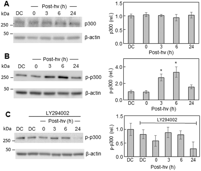
Fig. 4.
Total p300 and phospho-p300 levels before and after PDT: effects of a PI3K inhibitor. After pre-incubation with ALA, U87 cells were irradiated as described in Fig. 1. After the indicated periods of post-PDT incubation, cell samples were analyzed for total protein concentration, then subjected to Western blot analysis, using an antibody for overall p300 (A) or an antibody for p-p-300 (p-Ser-1834) (B). Other cells were treated with the PI3K inhibitor LY294002 (20 μM) immediately after irradiation, then checked for p-p300 levels at the indicated times (C). Dark controls (DC) −/+ LY294002 were analyzed as well. Total protein load per lane: 100 μg (A, B, C). Each immunoblot is a representative one from three replicate experiments; in each case, band intensities relative to β-actin and normalized to DC are plotted as means ± SEM (n=3); *P<0.01 vs. DC (plot B).

