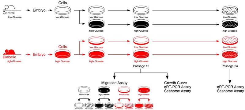
Figure 1. Schematic Depiction of Experimental Design.
Using the NOD mouse model, primary cells were isolated from embryos of normal and diabetic pregnancies. Cells were kept in culture medium with a glucose concentration designed to reflect the in vivo conditions of low glucose level (normal cells) and high glucose level (cells from diabetes-exposed embryos), respectively. After 6 Passages, half of each cell line was transferred into medium with the other glucose concentration, grown for 3 more Passages and frozen in multiple aliquots at Passage 9. For each assay, aliquots were thawed, and cells propagated in the respective medium until assay. In the migration assays, conditions of either high or low glucose in the assay were applied, resulting in 8 experimental groups.

