. 2019 Apr 30;28(2):183–215. doi: 10.5607/en.2019.28.2.183

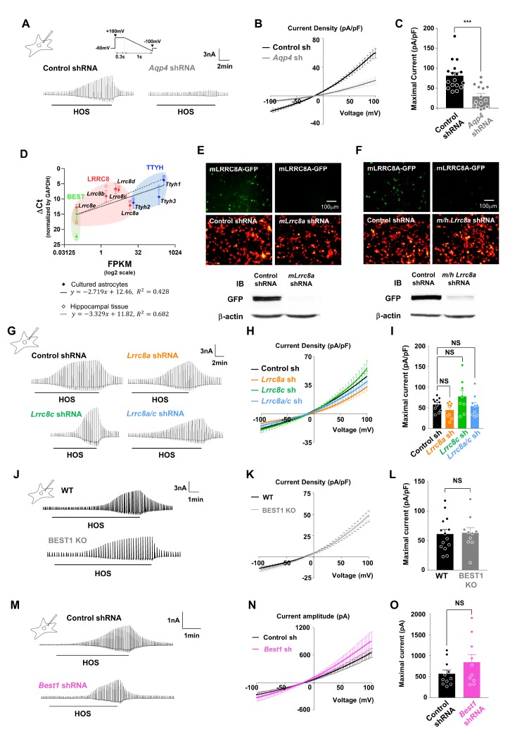
Fig. 1. Neither Lrrc8a nor Best1 encodes the pore-forming subunit of astrocytic VRACswell. (A) Representative HOS-induced current (ICl,swell) recordings from control shRNA and Aqp4 shRNA-expressing cultured astrocytes. Inset (left): schematic diagram of whole-cell patch-clamp recording in primary cultured astrocyte. Inset (right): schematic diagram of ramp protocol from +100mV to −100mV. (B) Averaged I~V curves for ICl,swell in control shRNA (black) and Aqp4 shRNA (grey). (C) Summary bar graph showing the maximal amplitude of ICl,swell from +100 mV to −100 mV. Data are represented as mean±SEM (n=17 for control shRNA and n=15 for Aqp4 shRNA; ***p<0.001, Mann-Whitney test). (D) Inverse correlation between the ΔCt value normalized by GAPDH measured by quantitative real-time quantitative PCR (qPCR) using cDNAs from hippocampal tissue (empty diamond), cultured cortical astrocytes (filled diamond) and FPKM value by RNA-sequencing that was previously shown in previous study (Zhang et al., 2014) to check relative mRNA expression of VRAC candidates such as Ttyh (Ttyh1, Ttyh2 and Ttyh3), Lrrc8 (Lrrc8a, Lrrc8b, Lrrc8c, Lrrc8d and Lrrc8e), and Best (Best1, Best2 and Best3) family. Data are represented as mean±SEM of ΔCt normalized by GAPDH (n=3 for cultured astrocytes and n=2 for hippocampal slices; R2=0.428 for cultured astrocytes and R2=0.682 for hippocampal slices; nonlinear regression). (E, F) Upper panels, fluorescence images of mLRRC8A-GFP and control or mLrrc8a or m/hLrrc8a shRNA-mcherry co-expressing HEK293T cells. Bottom panels, western blot results confirming the efficiency for mLrrc8a shRNA or m/h Lrrc8a shRNA compare to control shRNA in mLRRC8A-overexpressed HEK293T cells. (G) Representative ICl,swell recordings from control shRNA, Lrrc8a shRNA, Lrrc8c shRNA and both Lrrc8a and c shRNAs-expressing cultured astrocytes. (H) Averaged I~V curves for ICl,swell in control shRNA (black), Lrrc8a shRNA (orange), Lrrc8c shRNA (green) and both Lrrc8a and c shRNAs (sky blue). (I) Summary bar graph showing the maximal amplitude of ICl,swell from +100 mV to −100 mV. Data are represented as mean±SEM (n=11 for control shRNA, n=9 for Lrrc8a shRNA, n=8 for Lrrc8c shRNA, and n=9 for Lrrc8a/c shRNA; NS>0.05, ordinary one-way ANOVA). (J–L) Representative traces, averaged I~V curves and summary bar graphs of maximal ICl,swell currents from control or BEST1 KO mice in cultured astrocytes (n=14 for WT and n=10 for BEST1 KO; NS>0.05, Mann-Whitney test). (M~O) Representative traces, averaged I~V curves and summary bar graphs of maximal ICl,swell currents from control (n=11) or BEST1 shRNA expressing condition (n=10) in cultured astrocytes (n=11 for control shRNA and n=10 for Best1 shRNA; NS: p>0.05, Two-tailed unpaired t-test).

Fig. 1