. 2019 Apr 30;28(2):183–215. doi: 10.5607/en.2019.28.2.183

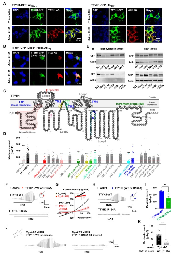
Fig. 8. R165 in TTYH1 is the pore-forming amino acid as evidenced by mutagenesis-based structure-activity analysis. (A, B) Non-permeabilized (−Triton X-100) and permeabilized (+Triton X-100) HEK293T cells expressing TTYH1-GFP were immunostained with N terminus of TTYH1 by anti-TTYH1 antibody, C terminus of TTYH1 by anti-GFP antibody and Loop1-FLAG-tagged-TTYH1 clone by anti-FLAG-tag antibody. (C) The prediction of membrane topology of TTYH1 based on permeabilization staining results and topology prediction software (TMHMM). Epitope for AbN-term antibody (red box), Δ2A4 (H131-T135, pink circle), Δ2C2 (V161-R165, yellow-green circle), Δ2C3 (M166-A170, green circle), Δ4B(L285-N304, sky blue), ΔYY (Y299-Y300, blue circle) and R165 (which was the pore of TTYH1) were also indicated. (D) Summary bar graph showing the maximal amplitude of ICl,swell from +100 mV to −100 mV recorded with WT or several truncation mutants of TTYH1 co-expressing HEK293T cells. Data are represented as mean±SEM (n=22 for AQP4 expressing HEK293T cells, n=57 for WT-TTYH1 with AQP4 co-expressing HEK293T cells, n=9 for loop1-TTYH1, n=7, 8, 11, 8 and 15 for related to loop2A-TTYH1, n=9 for loop2B-TTYH1, n=8, 6, 9, 10, 16, 11 for related to loop2C-TTYH1, n=7 for both loop2D and loop2E-TTYH1, n=8 for loop4A, n=8 and 15 for related to loop4B-TTYH1, and n=9, 11, 8, 9 and 8 for loop4C, D, E, F and G of TTYH1; **<0.01, ***<0.001, ****<0.0001, Kruskal-Wallis test). (E) Surface expression of truncation mutants of TTYH1 that showed significant reduction of ICl,swell by biotinylation assay. (F, G) Representative traces and averaged I~V curve for ICl,swell from wild-type (WT) and R165A-TTYH1 with AQP4 co-expressing HEK293T cells. (H) Representative traces for ICl,swell from TTYH2-R164A with AQP4 compared to TTYH2-WT with AQP4 overexpressing CHO cells. Inset: schematic diagram of whole-cell patch-clamp recording in R164A or WT-TTYH2 with AQP4 expressing CHO cells. (I) Summary bar graph showing the maximal amplitude of ICl,swell from −100mV to 100mV. Date are represented as mean±SEM (n=4 and 9 for TTYH2-WT with AQP4 and TTYH2-R164A with AQP4 coexpressing HEK293T cells, **p<0.01; Mann-Whitney test). (J) Representative ICl,swell recordings from Ttyh 1/2/3 shRNAs with shRNA-insensitive clone of TTYH1-WT and Ttyh1/2/3 shRNAs with shRNA-insensitive clone of TTYH1-R165A-expressing cultured astrocytes. Inset: schematic diagram of whole-cell patch-clamp recording in primary cultured astrocyte. (K) Summary bar graph showing the maximal amplitude of ICl,swell from +100 mV to −100 mV in the presence of the Ttyh 1/2/3 shRNAs with shRNA-insensitive clone of TTYH1-WT (Ttyh1 sh-insens.) or with shRNA-insensitive clone of TTYH1-R165A-expressing cultured astrocytes. Data are represented as mean±SEM (n=18 and 10 for Ttyh1/2/3 shRNAs with shRNA-insensitive form of WT-TTYH1 and R165A-TTYH1 expressing astrocytes, respectively; ****<0.0001, unpaired t-test).

Fig. 8