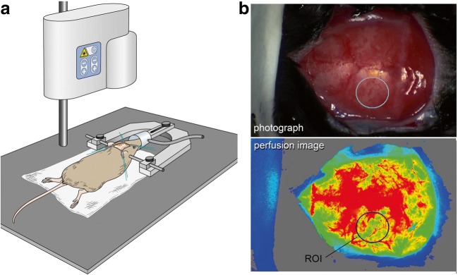
Fig. 1.
Determination of cerebral cortical perfusion. a The perfusion measurement: The animal is mounted on a stereotaxic frame. After the skin incision, the laser SPECKLE camera is placed over the animal to acquire perfusion images. b The evaluation of cerebral perfusion (upper image: photograph, lower image: flux image visualizing cerebral cortical perfusion). A region of interest of 7 mm2 is placed on the left parietal region to measure perfusion flux values

