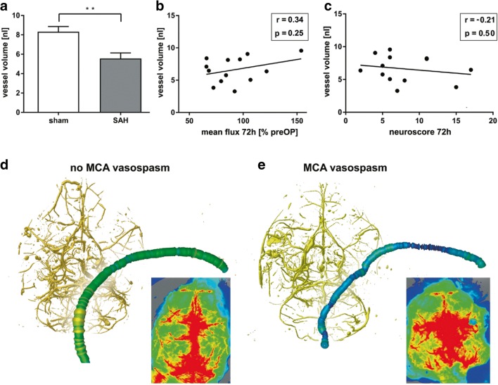
Fig. 4.
CV and cerebral perfusion after SAH. a Vessel volumes of a 2.5-mm segment of the MCA distal of the carotid T after SAH and sham surgery. Note that the vessel volumes are significantly lower in SAH animals, indicating CV. b, c The correlation of MCA vessel volume with perfusion and neuroscore. d, e Exemplarily show the reconstructed vascular tree and cerebral perfusion in a sample without CV (d) and with CV (e), in which CV was not associated with impaired perfusion

