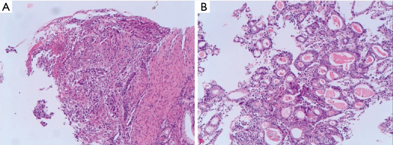
Figure 1.
Pathological section used for diagnosis. (A,B) Hematoxylin and eosin staining, magnification, 200×: the tumor was moderately differentiated adenocarcinoma (the moderately differentiated regions accounted for approximately 70% and the poorly differentiated regions accounted for approximately 30% of the tumor).

