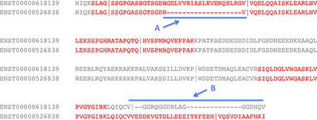
Box 2, Figure I. Identifying alternative splice events.
Part of an alignment between two splice isoforms of the gene EEF1D. Identified peptides are in red font and vertical lines mark the position of exon boundaries. The two regions that distinguish the isoforms are marked as A and B and the extent of the differences between the two regions are marked by a blue line. Region A differs by an indel; peptides that map to both sides of the indel confirm the translation of this splice isoform. By contrast, peptides map to just one side of the splice event in region B (a C-terminal substitution), so the translation of an alternative isoform with the alternative C terminus is not confirmed.