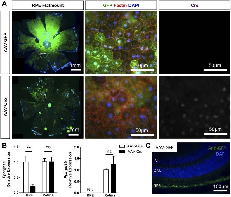
Figure 4. RPE-specific deletion of PGC-1 isoforms in adult mice.
(A) RPE/choroid flat-mounted preparations from adult PGC-1α;PGC-1β–double floxed mice 1 mo after subretinal injection with AAV-GFP and AAV-Cre. Immunolocalization of the GFP reporter (green) confirmed efficient viral transduction of the entire RPE surface. High magnifications in the mid-periphery (dotted squares) show GFP+ RPE cells labeled with F-actin (red). Cre expression (white) was only detected in the nuclei of RPE from AAV-Cre–injected animals. (B) PGC-1α and PGC-1β gene expression analysis in RPE/choroid and retinal tissues from AAV-GFP and AAV-Cre mice 1 mo postinjection (n = 3–6). (C) Representative ocular cryosection from experimental mice 4 mo after AAV-GFP injection, showing highly selective viral transduction of the RPE cells. Scale bar is 100 μm. Error bars are means ± SEM. Data were analyzed by unpaired t test. **P ≤ 0.01 compared with their respective AAV-GFP control mice.

