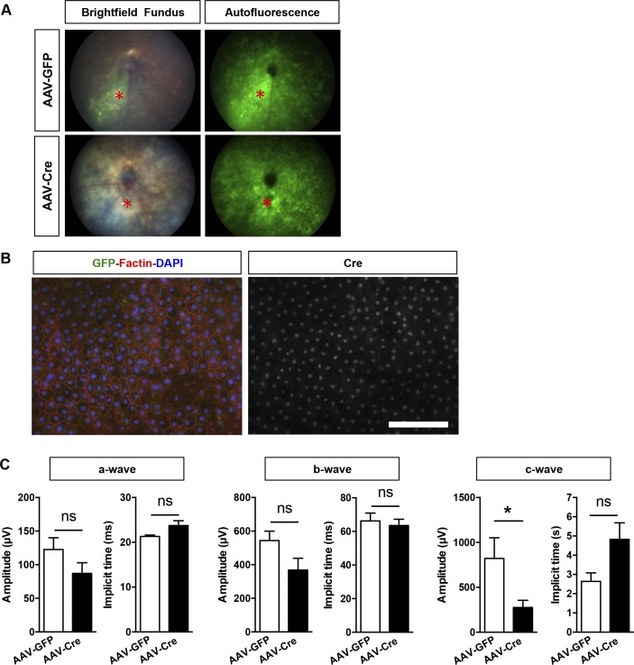
Figure S3. Morphological and functional evaluation of adult PGC-1–floxed mice 2 and 4 wk after subretinal AAV injection.
(A) Example of fundus imaging GFP fluorescence detection confirming the pan-retinal transduction of the AAVs 2 wk after subretinal injection in experimental floxed animals. The asterisk (*, red) indicates the site of injection. (B) RPE/choroid flat mounts immunostained for F-actin (red) and Cre (white) from AAV-Cre–injected experimental mice revealing Cre+ nuclei in a large area of the epithelium 1 mo postinjection. No major morphological changes were observed. Scale bar is 100 μm. (C) ERG analysis of the a-, b-, and c-wave amplitudes and implicit times 1 mo postinjection (n = 5–6). Error bars are means ± SEM. Unpaired t tests were used in all comparisons. *P ≤ 0.05 compared with their respective AAV-GFP mice.

