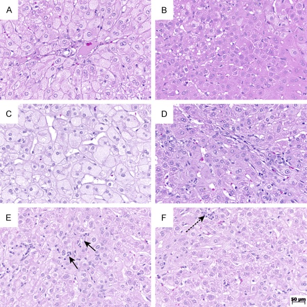
Figure 1.

Hematoxylin and eosin stain at original magnification, 200×. A. Rabbit from placebo group with high score of ballooning; B. Liver from the ramipril group with preserved lobular architecture, showing no ballooning; C. Rabbit from placebo group with high score of steatosis; D. Rabbit from the ramipril group with minor degree of steatosis; E. Rabbit from the placebo group with high score of lobular inflammation; F. Rabbit from the ramipril group with score 1 of lobular inflammation.
