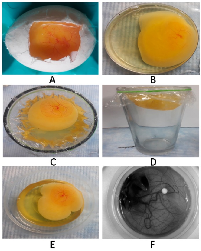
Figure 1.
Representative images of chorioallantoic membrane (CAM) variants. (A) in-ovo setup by windowing method on day of incubation; (B) ex-ovo setup in a Petri plate; (C) ex-ovo setup in a glass-vertical view; (D) ex-ovo setup in a glass-horizontal view; (E) ex-ovo setup on plastic cups, image taken by a Camera and; (F) ex-ovo setup in plastic cups, image taken by a Chemidoc (Charge-coupled device (CCD) Camera).

