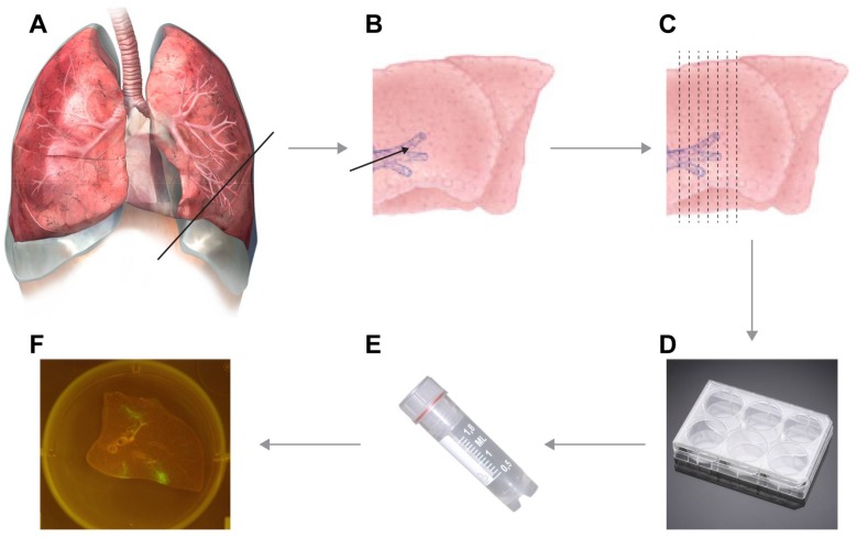
Figure 1.
Flow scheme of the experimental design. Lungs are resected during necropsy (A) and inflated with warm liquid low-melting point agarose (B). After solidification on ice, thin slices of approximately 1-mm thick can be cut by hand (C) and are transferred to 6- or 24-well plates pre-filled with culture medium (D). Slices can subsequently be inoculated with the desired virus (E) and infection of the slices is followed in time (F). In this example in panel F, a macaque lung slice infected with recombinant measles virus is shown.

