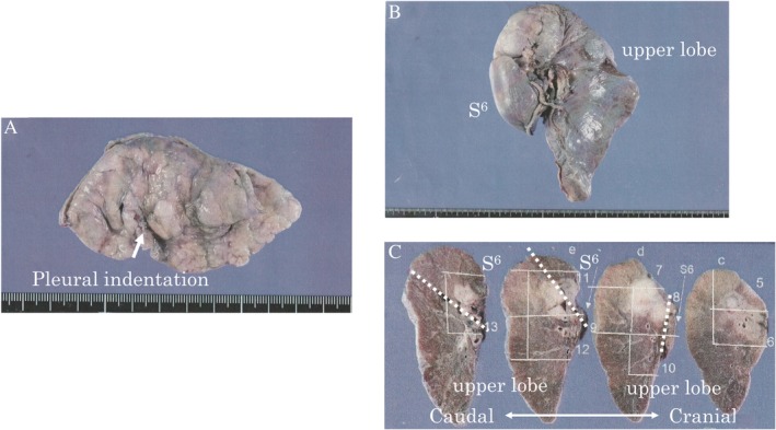
Figure 3.

Macroscopic findings of the resected specimens. (A) Partial resection of the right lower lobe. Pleural indentation by the right segment (S) 9 tumour was identical, but the visceral pleura was intact. (B, C) resection of the upper left lobe together with the S6 area. invasion of the left S1 + 2 tumour into the S6 area was observed but not exposed externally or disseminated. the white dotted line indicates the fissure line between the upper and lower lobes.
