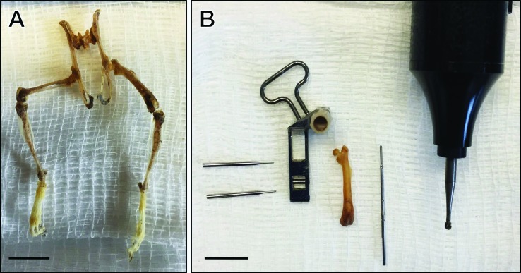
Figure 3.
Dermestid beetle–cleaned skeletons for training surgical techniques. (A) Dermestid beetle–cleaned skeleton allows visualization of the size and scale of the surgical area and skeletal anatomy. (B) Equipment for LockingMouseNail (RISystem, Davos, Switzerland) surgery alongside an isolated left femur gives trainee an idea of the size ratio between surgical implants and femur. From left to right: locking pins, locking nail guide arm, mouse femur, locking nail, and microdrill with 1.6-mm burr. Scale bar, 1 cm.

