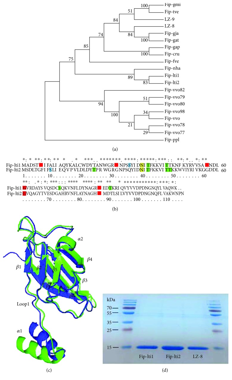
Figure 1.
Phylogenetic and structural features and expression of Fip-lti1 and Fip-lti2. (a) Phylogenetic analysis of FIPs. (b) Sequence alignment of Fip-lti1 and Fip-lti2. Specific posttranslational modification sites are indicated as colored amino acids: myristoylation (red), casein kinase II phosphorylation (blue), protein kinase C phosphorylation (green), and N-glycosylation (yellow). (c) Superposition of the main chain backbone of Fip-lti1 (green) with Fip-lti2 (blue). (d) SDS-PAGE analysis of purified FIPs.

