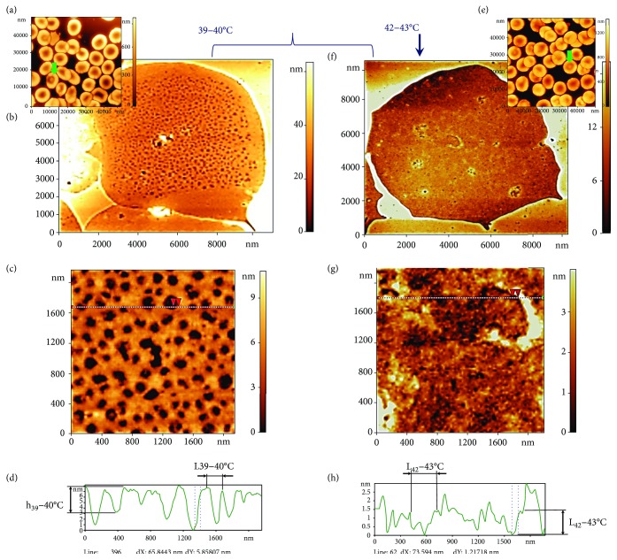
Figure 6.
Influence of temperature at 39–40°С and 42–43°С on spectrin matrix nanostructure. (a, e) AFM 3D images of RBC morphology, 38 × 38 μm2 and 60 × 60 μm2. (b, f) AFM 2D images of the spectrin matrix, 12 × 12 μm2. (c, g) AFM 2D images of a fragment of the spectrin matrix nanostructure, 2000 × 2000 nm2. (d) Height profile along the lines indicated in (c, h, g). Red arrows correspond to dashed lines on the profile. The curly bracket unites possible variants of RBC ghosts and spectrin matrix at 39–40°С. The vertical arrow shows a ghost RBC variant and the spectrin matrix at 42–43°С. Ensemble 1 of RBC ghosts corresponds to (b–d). Ensemble 2 of RBC ghosts corresponds to (f–h).

