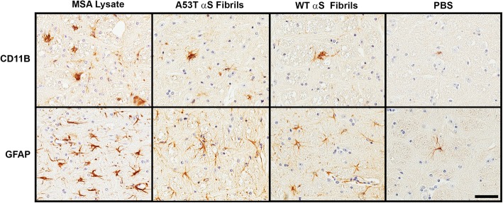
Fig. 4.
Representative immunohistochemistry of microglia and astrocytes in M83+/− mice neonatally injected at P0 with PBS, WT human αS fibrils, A53T human αS fibrils or MSA brain lysates. M83+/− mice were injected at P0 as described in “Material and Methods” and aged. Images showing microgliosis with an anti-CD11B antibody and astrogliosis with an anti-GFAP antibody in the pons of mice with induced αS pathology. Scale bar = 50 μm

