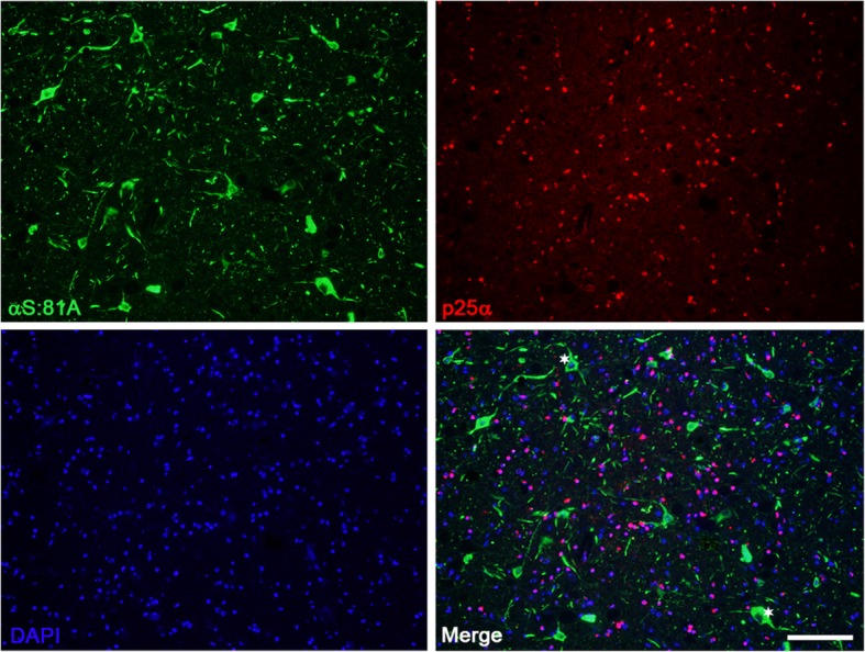
Fig. 7.

Immunofluorescence analysis demonstrating the paucity of αS pathology within oligodendrocytes of M83+/− mice treated with MSA lysates. M83+/− mice that developed αS pathology following injection with MSA derived cerebellar lysates were evaluated through double immunofluorescence for the cell type within which pathology formed. αS pathology identified with antibody 81A was not seen to co-localize with oligodendroglial marker p25α. Asterisks indicate αS pathology in neuronal bodies. Scale bar = 100 μm
